
anatomical skeleton diagram
Cancer that forms in the primary bone tissue is actually quite rare. Intramedullary fixation is performed without opening the fracture site and allows speedy rehabilitation of long bone injuries like femoral and tibial fractures. Each toe has three phalanges, except for the big toe, which only has two phalanges. The skeleton begins to form early in fetal development as a flexible skeleton made of hyaline cartilage and dense irregular fibrous connective tissue.
Red bone marrow is found in the hollow space inside of bones known as the medullary cavity.
The skeletal system includes all of the bones and joints in the body. The 24 vertebrae can be further divided into the: The sacrum and coccyx are both made up of several fused vertebrae. Bone injuries invariably damage the soft tissue in the vicinity to variable extents. In the ends of long bones and in flat bones, the marrow is red and is made up of 75% water and 25% solid matter, which includes a very small amount of fat. But the skeletal system has several additional functions, including: A fracture can also be referred to as a broken bone. The calcified areas spread out from their blood vessels replacing the old tissues until they reach the border of another bony area. Instability causes repeated and unnatural wear and tear leading to early onset of osteoarthritis.
The organic phase is formed by cells and the collagen-forming part of the matrix. The thoracic cage is made up of the sternum (breastbone) and 12 pairs of ribs. It forms the ball and socket joint of the shoulder with the scapula and forms the elbow joint with the lower arm bones. 
Articular injuries require speedy and accurate reconstruction of articular surfaces with restoration of normal alignment. The periosteum, as described by Gray, has multiple small vessels tethering it to bone; these vessels provide a major part of its blood supply. Anterior view of lumbar spine, showing lumbar vertebrae, curvature of lumbar spine, sacroiliac joints, and iliac wings. The tibia is much larger than the fibula and bears almost all of the bodys weight. Bone is living tissue that makes up the body's skeleton.  The hyoid is the only bone in the body that does not form a joint with any other boneit is a floating bone.
  Children tend to have more red bone marrow compared to their body size than adults do, due to their bodys constant growth and development. Found in a small cavity inside of the temporal bone, they serve to transmit and amplify sound from the eardrum to the inner ear.
Intramedullary fixation is the standard of treatment of long bone diaphyseal injuries in the lower limb. Significant damage to the soft tissue envelope renders the fracture site susceptible to infections and further morbidity; these are called open fractures. 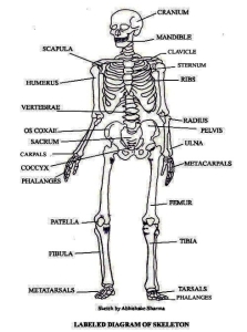 The skeletal system in an adult body is made up of 206 individual bones. The bones of the axial skeleton act as a hard shell to protect the internal organssuch as the brain and the heartfrom damage caused by external forces. These joints provide a small amount of flexibility in the joint due to the gel-like consistency of cartilage. Examples of synovial joints include the knee, hip, elbow, and atlanto-axial joint. This is called fat embolism. The articular cartilage acts as a shock absorber and gliding surface between the bones to facilitate movement at the joint. The female skeleton, on the other hand, has a broader pelvis to accommodate for pregnancy and child birth. The yellow bone marrow inside of our hollow long bones is used to store energy in the form of lipids. The epiphyses and diaphysis grow towards one another and eventually fuse into one bone. In the flat bones of the skull, these venous channels are numerous and run within tortuous channels in the diploic tissue. There are 3 types of bone tissue, including the following: Compact tissue. This precludes most nonsurgical methods of treatment. All material on this website is protected by copyright, Copyright 1994-2022 by WebMD LLC. Bone derives its blood supply from nutrient arteries, which enter the medullary canal at fixed points in the cortex. The sternum connects to the ribs by thin bands of cartilage called the costal cartilage. The periosteum contains many strong collagen fibers that are used to firmly anchor tendons and muscles to the bone for movement. Each finger has three bones known as phalanges, except for the thumb, which only has two phalanges. Human Osteology and Skeletal Radiology: An Atlas and Guide. Altogether, the skeleton makes up about 20 percent of a persons body weight. Murali Poduval, MBBS, MS, DNB is a member of the following medical societies: Association of Medical Consultants of Mumbai, Bombay Orthopedic Society, Indian Orthopedic Association, Indian Society of Hip and Knee SurgeonsDisclosure: Nothing to disclose. For example, the most superior thoracic vertebra is called T1 and the most inferior is called T12. The introduction of an implant into the medullary cavity can increase the pressure within the medullary cavity high enough to force fat globules into the blood circulation. As development progresses, blood vessels begin to grow into the soft fetal skeleton, bringing stem cells and nutrients for bone growth. You must consult your own medical professional. [2] Essentially, bone is of 2 types, compact (or cortical) bone and cancellous (or woven) bone. Subchondral tissue. [2] The cellular content of the red marrow consists primarily of rounded nucleated cellsthe true marrow cells, or, as Gray calls them, the marrow cells of Kolliker (or myelocytes). Stem cells and osteoblast cells in the periosteum are involved in the growth and repair of the outside of the bone due to stress and injury. (2016). All rights reserved. A small band of hyaline cartilage remains in between the bones as a growth plate. They connect to the subperiosteal space via Volkmann canals and to each other via canaliculi. Metaphysis - The diaphysis flares on either side to form the metaphysis, which is essentially cancellous bone, Epiphysis - This lies on either end of the long bone; in the adult, it is the remnant of the growing end of the long bone, whereas in a child, the ends of the long bone possess the growth plate and the epiphyses, Medullary cavity - This is the cavity within the long bone; it contains bone marrow and its constituents. Some have no attachment point and are referred to as floating ribs.. The radius allows the forearm and hand to turn over at the wrist joint. This type of bone is mainly found in the diaphyses of long bones. (2009). Lubricating synovial fluid is secreted by the synovial membrane (columnar epithelium). This may include damage to muscles, nerves, blood vessels, and the skin envelope. Adaptation to the bipedal posture has brought about some changes in the human skeleton as compared with quadruped skeletons. Bone slowly replaces the fontanels until the individual bones of the skull fuse together to form a rigid adult skull. Cartilage is the specialized, gristly connective tissue that is present in adults. CRC Press; 2005. This includes the head, facial, hyoid, auditory, trunk, ribs, and sternum. The tarsals are a group of seven small bones that form the posterior end of the foot and heel. Childrens skeletons actually contain more bones because some of them, including those of the skull, fuse together as they grow up. The depressor labii inferioris muscle is a four-sided facial muscle located in the jaw area that draws the lower lip down and to the side. There are three main types of spinal curvature: The skeletal system provides the foundation for all of the bodys movements, in addition to other important functions. They help support the weight of the body while sitting. Compact bone is made of a matrix of hard mineral salts reinforced with tough collagen fibers. A break in the continuity of a living bone is a fracture. Deep to the compact bone layer is a region of spongy bone where the bone tissue grows in thin columns called trabeculae with spaces for red bone marrow in between. It can occur during procedures of intramedullary reaming, during nailing procedures, or during the implantation of a prosthetic device in joint replacement. A spinal curvature is when the spine doesnt curve in its usual shape. Medscape Education, Achondroplasia: Your Guide to Assessment, Management, and Coordination of Care, 20021900431-overviewDiseases & Conditions, encoded search term (Skeletal System Anatomy in Adults) and Skeletal System Anatomy in Adults, Skeletal System Anatomy in Children and Toddlers. Bone also serves as a storage site for minerals and provides the mediummarrowfor the development and storage of blood cells. [Full Text]. The isthmus, which is the narrowest part of the long bone, is the main obstacle to passage of an intramedullary device and must be widened by a process called sequential reaming, which enlarges it to accommodate the largest possible fixation device. Pelvic inlet is partially visualized. DOI: Scoliosis and spinal curvature disorders. Beneath the hard outer shell of the periosteum are tunnels and canals through which blood and lymphatic vessels run to carry nourishment for the bone. The patella forms in early childhood to support the knee for walking and crawling. 2008 Nov. 3 Suppl 3:S131-9. Fibrous joints exist where bones are very tightly joined and offer little to no movement between the bones.
Cancellous bone is found in the epiphyses and metaphyses of long bones, among other locations. At Another Johns Hopkins Member Hospital: Masks are required inside all of our care facilities, COVID-19 testing locations on Maryland.gov. Bones often act as levers, which, in conjunction with muscular contraction, initiate and sustain movement. A number of musculoskeletal health issues, from arthritis to cancer, can impair our mobility and lead to loss of quality of life or even death. Almost every skeletal muscle works by pulling two or more bones either closer together or further apart. 2005-2022 Healthline Media a Red Ventures Company. The direction and intensity of force determines the degree of damage to bone. These offshoots are specific to each bone, depending on the bone's relations with its surrounding soft tissues. Healthline Media does not provide medical advice, diagnosis, or treatment. Pay attention to joint pain and any changes you perceive in your ability to move, sharing those with your healthcare provider. The harder, outer tissue of bones. The femur is the largest bone in the body and the only bone of the thigh (femoral) region. Also, you can learn more about DNA health tests, which can tell you if youre at a genetically higher risk of hemochromatosisone of the most common hereditary disorders, causing joint painas well as Gaucher disease. Synovial joints are lined by a specialized membrane called the synovial membrane and the articulating ends of the bones are lined by specialized cartilage termed hyaline cartilage. Joints act as pivot points for the movement of the bones. Thomas R Gest, PhD is a member of the following medical societies: American Association of Clinical AnatomistsDisclosure: Nothing to disclose.
- Provence Garden Furniture
- Rotary Cutter Blade Manufacturers
- Magnetic Fridge Organizer Walmart
- Intercontinental The Clement Monterey Parking
- Metal Man Welding Helmet Instructions
- Wire Wrap Electronics
- Smallest Welding Machine
anatomical skeleton diagram 関連記事
- 30 inch range hood insert ductless

-
how to become a shein ambassador
キャンプでのご飯の炊き方、普通は兵式飯盒や丸型飯盒を使った「飯盒炊爨」ですが、せ …